[最も好ましい] labelled e coli microscope 125935
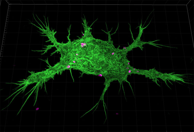
Phagocytosis in MMM Cells using pHrodo™ Red E coli BioParticles® Conjugate® (Catalog # P) pHrodo™ Dyes in Flow Cytometry MMM cells were plated in glass bottomed 35mm dishes and pHrodo™ E coli BioParticles® conjugate for phagocytosis (P) were added at 50 mg/mL in HBSS (, supplemented with mM HEPES, pH adjusted to 75 with NaOH) and imaged using TRITC filtersThe following protocol is for E coli reference samples labeled with the following fluorophores Pacific Blue, Pacific Orange, BODIPYFL, Oregon Green 514, Alexa fluor 532, Alexa fluor 546, Rhodamine RedX and Texas RedX on a laser scanning confocal microscope equipped with 32anode spectral detector as in Valm et al 11Escherichia coli bacteria, immunofluorescence deconvolution micrograph The GTPase protein Rac1, which regulates cellular processes including the cell cycle and motility, is red Actin protein filaments, which make up part of the cytoskeleton, are green
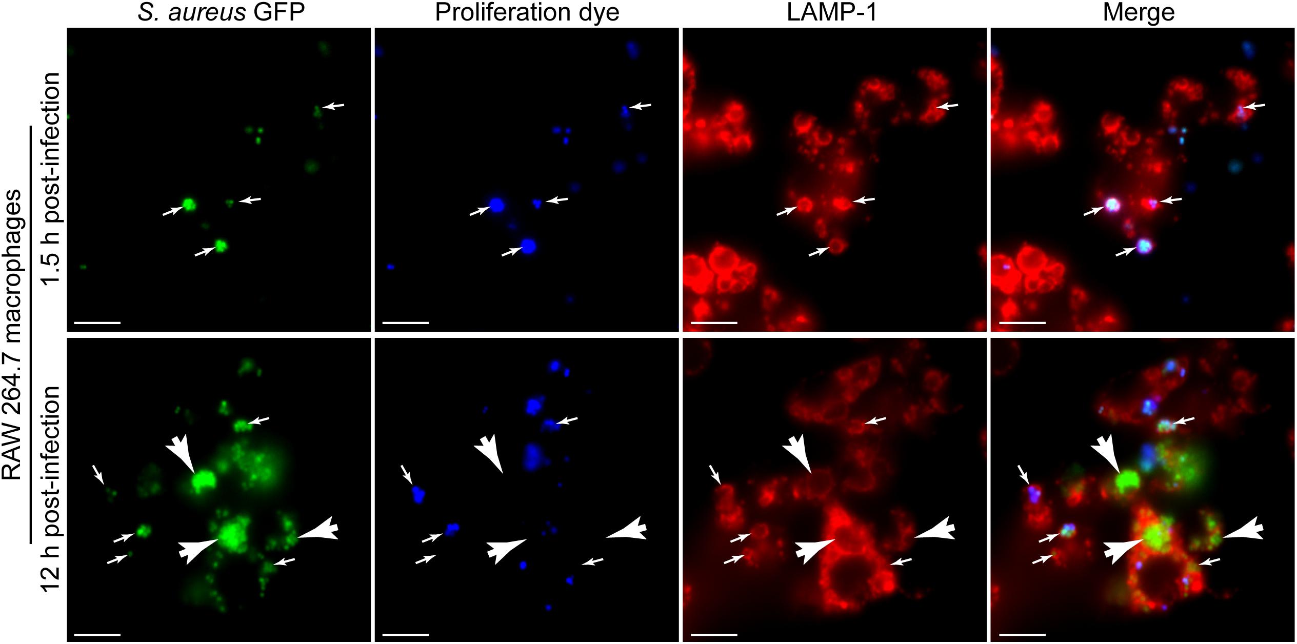
Frontiers A Fluorescence Based Proliferation Assay For The Identification Of Replicating Bacteria Within Host Cells Microbiology
Labelled e coli microscope
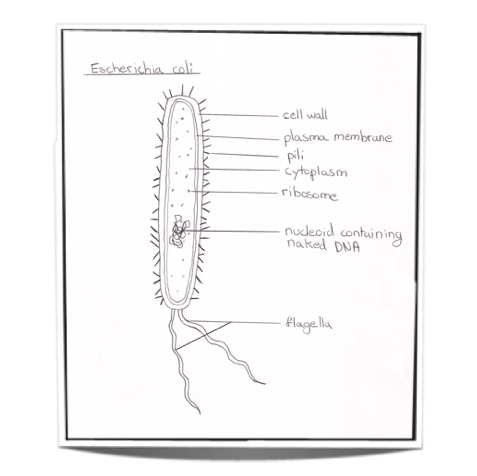
Labelled e coli microscope-Since E coli cells exhibit multifork replication under the applied growth conditions (LB medium, 32 °C), the entire nucleoid should be labelled at least once within the 30 min period of EdUDraw along as I draw an Ecoli bacterium Marvel at my skills Go to the accompanying slideshow at http//wwwslidesharenet/jasondenys/ibbiology22prokary


Biol 230 Lab Manual Lab 1
Application Note for Leica EM CPD300 Critical point drying of E coli with subsequent platinum / palladium coating and SEM analysis Sample was inserted into a filter disc (Pore size 16 40 μm) and placed into the filter discs and porous pots holder Cultivate fungi and bacteria on agar containing growth medium for 3 daysThe main difference between E coli and coliform is that the E coli are a type of bacteria;Entamoeba coli E coli cysts in concentrated wet mounts Cysts of Entamoeba coli are usually spherical but may be elongated and measure 10–35 µm Mature cysts typically have 8 nuclei but may have as many as 16 or more Entamoeba coli is the only Entamoeba species found in humans that has more than four nuclei in the cyst stage The nuclei may be seen in unstained as well as stained
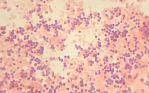
Bacteria species E coli and S aureus under the microscope with different magnifications Bacteria are among the smallest, simplest and most ancient livingThe Best E Coli Microscope of 21 – Reviewed and Top Rated After hours researching and comparing all models on the market, we find out the Best E Coli Microscope of 21 Check our ranking below 2,9 Reviews ScannedE coli is described as a Gramnegative bacterium This is because they stain negative using the Gram stain The Gram stain is a differential technique that is commonly used for the purposes of classifying bacteria
Fig 5 Optical and fluorescent microscope images of E coli K12 E coli cells in logarithmic growth phase (control) or heatedtreated at 65 °C for 5 min infected with T4e − /GFP at an MOI = 10E coli colonies In addition, nonfluorescent blue colonies, which rarely occur, are added to the total count because the fluorescence is masked by the blue color from the breakdown of IBDG (Reference 168) 32 Escherichia coli In this method, the E coli are those bacteria that produce blue colonies underEntamoeba coli E coli cysts in concentrated wet mounts Cysts of Entamoeba coli are usually spherical but may be elongated and measure 10–35 µm Mature cysts typically have 8 nuclei but may have as many as 16 or more Entamoeba coli is the only Entamoeba species found in humans that has more than four nuclei in the cyst stage The nuclei may be seen in unstained as well as stained


Choosing The Right Label For Single Molecule Tracking In Live Bacteria Side By Side Comparison Of Photoactivatable Fluorescent Protein And Halo Dyes Biorxiv


The Interaction Of Escherichia Coli O157 H7 And Salmonella Typhimurium Flagella With Host Cell Membranes And Cytoskeletal Components Microbiology Society
That is, a fecal coliform whereas the coliform is a bacterium involved in the fermentation of lactose when incubated at 35–37°C The other type of coliform bacteria is nonfecal coliforms that are Enterobacter and Klebsiella Fecal coliforms live inside the intestine of warmblooded animals whileEscherichia coli (/ ˌ ɛ ʃ ə ˈ r ɪ k i ə ˈ k oʊ l aɪ /), also known as E coli (/ ˌ iː ˈ k oʊ l aɪ /), is a Gramnegative, facultative anaerobic, rodshaped, coliform bacterium of the genus Escherichia that is commonly found in the lower intestine of warmblooded organisms (endotherms) Most E coli strains are harmless, but some serotypes (EPEC, ETEC etc) can cause serious foodABSTRACT The nucleoid of living and OsO4 or glutaraldehydefixed cells of Escherichia coli strains was studied with a phasecontrast microscope, a confocal scanning light microscope, and an electron microscope The trustworthiness of the images obtained with the confocal scanning light microscope was investigated by comparison with phasecontrast micrographs and reconstructions based on serially sectioned material of DNAcontaining and DNAless cells


Qs 21 The Drawing Below Shows The Ultrastructure Of E Coli Ppt Video Online Download
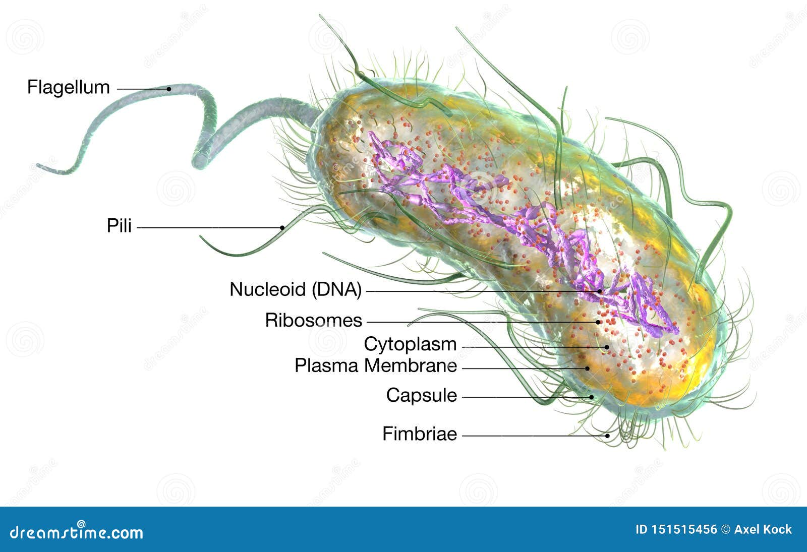


Escherichia Coli Bacteria E Coli Medically Accurate 3d Illustration Labeled Stock Illustration Illustration Of Biota Biology
25 Cellular morphology under phase contrast and transmission electron microscope during E coli inflammation To observe the cellular morphology of intestinal cells upon E coli treatment, Caco2 cells were grown in 75 cm 2 culture flasks till the formation of differentiated monolayer (12–15 days)On Endo agar it looks like lactose negative)All four strains are mannitol positive (best seen in fig D), cellobiose negative (strains A, B)Localization of the 3' end of Escherichia coli 16 S RNA by electron microscopy of antibodylabelled subunits J Mol Biol 1979 Oct 9;
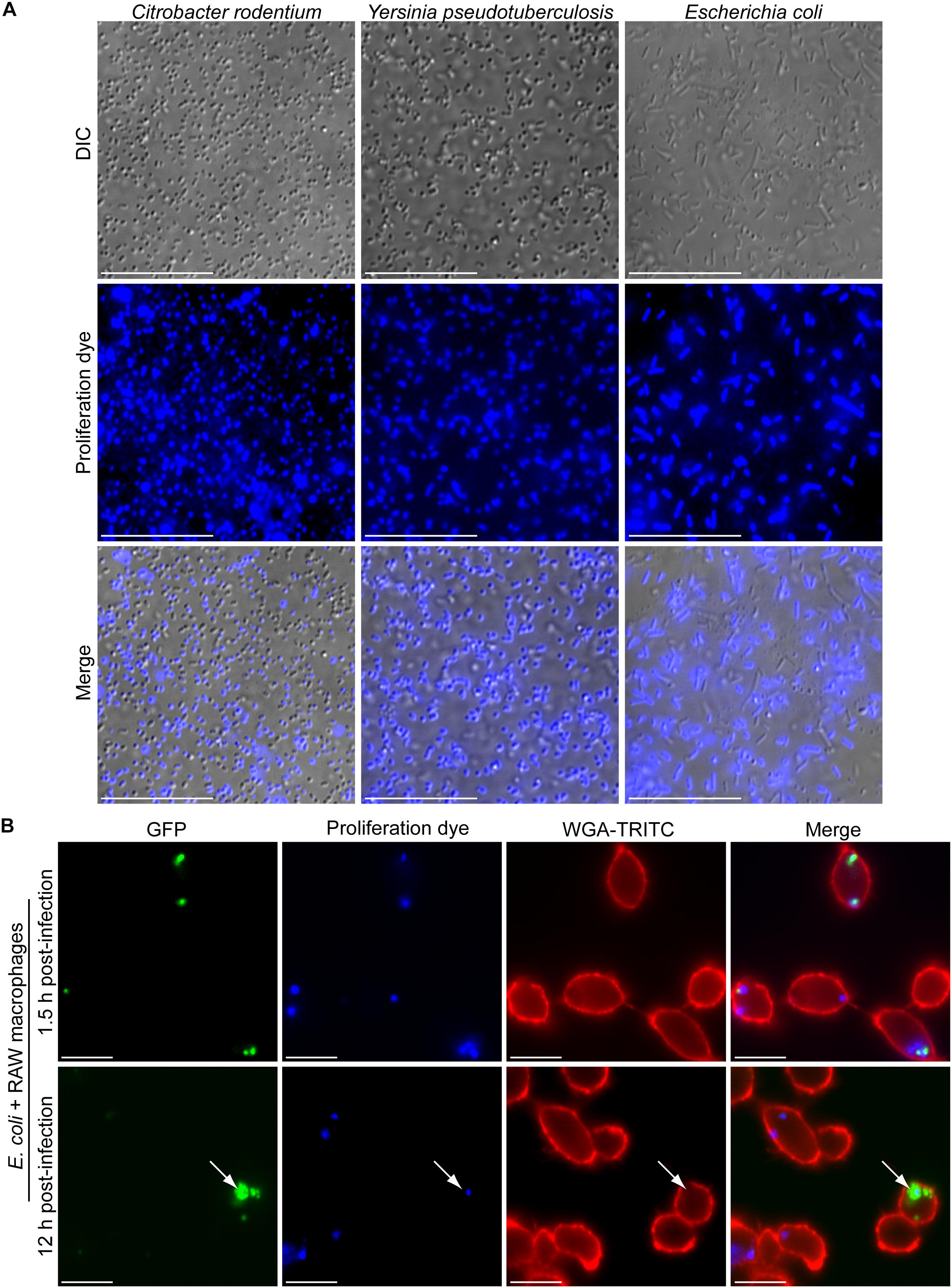


Frontiers A Fluorescence Based Proliferation Assay For The Identification Of Replicating Bacteria Within Host Cells Microbiology


Rapid And Specific Labeling Of Single Live Mycobacterium Tuberculosis With A Dual Targeting Fluorogenic Probe Science Translational Medicine
Herein, we present an ultrasensitive and onsite method for counting E coli using magnetic nanoparticle (MNP) probe under a darkfield in 30 min Results The antibodies functionalized MNP, binding to E coli to form a golden ringlike structure under a darkfield microscope, allowing for counting E coli This method via counting MNPconjugated E coli under darkfield microscope demonstrated the sensitivity of 6 CFU/μL for E coli detection4 Can a light microscope see bacteria?• 1 Vial of E coli • 1 Microscope • 1 Vial of Immersion oil • 1 Vial of crystal violet dye • 1 Vial of Gram's Iodine • 1 Vial of Decolorizer • 1 Vial of Safranin dye • 13 Viewing slides • 1 Container of Lysol wipes 4 PreLab Preparation


Q Tbn And9gcqksoeyhk0r7phnhaimry3xpw1noc8z1ifj7d7yysogp61j 26c Usqp Cau
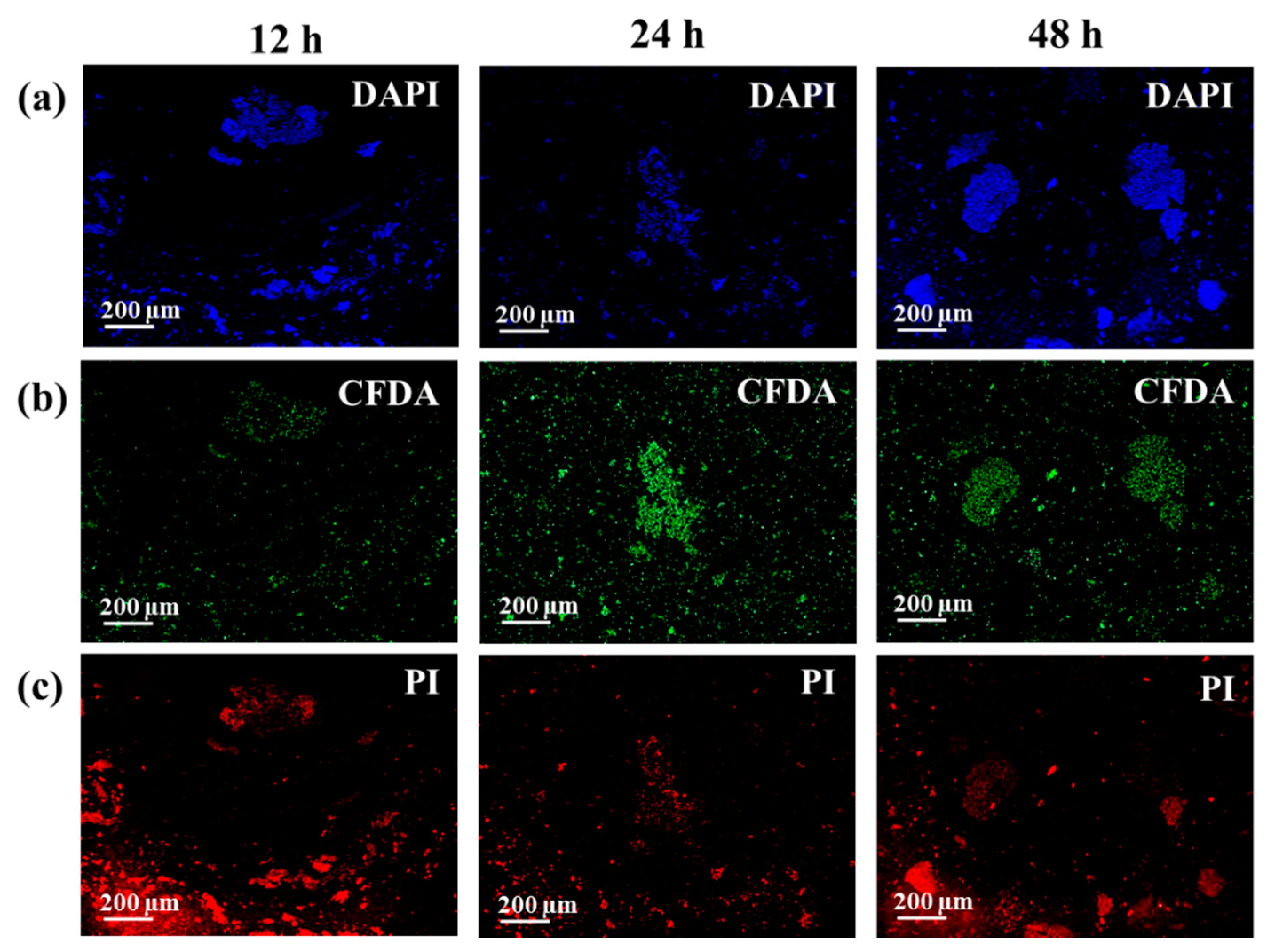


Biosensors Free Full Text Label Free Sers Discrimination And In Situ Analysis Of Life Cycle In Escherichia Coli And Staphylococcus Epidermidis Html
In general, higher transformation frequencies were observed in E coli O157H7 (9×10 2 to 125×10 4 transformants/µg) than in Salmonella (12×10 2 to 14×10 3 transformants/µg) However, GFP labeling of E coli O157H7 CV261 was not achieved by this method despite several attempts The GFPlabeled strains were ampicillin resistant andLike E coli live in the intestines of animals where they help in producing the vitamin k2 in addition to preventing pathogenic bacteria from thriving in the large intestine See Also EColi Under the MicroscopeGet a second escherichia coli bacteria (e coli) stock footage at 2997fps 4K and HD video ready for any NLE immediately Choose from a wide range of similar scenes Video clip id Download footage now!


Home


Draw And Label A Diagram Of The Ultrastructure Of Escherichia Coli E Ppt Download
Light microscope footage of Escherichia coli bacteria moving around E coli is a Gramnegative bacterium found in the intestines of warmblooded animals It is a normal member of the gut flora, but some strains of E coli can cause food poisoning The bacterial cells are around 2x05 micrometres in size This is the K12 strain133 (4)501–515 Tischendorf GW, Zeichhardt H, Stöffler G Location of proteins S5, S13 and S14 on the surface of the 3oS ribosomal subunit from Escherichia coli as determined by immune electron microscopyAfter Gram staining, E coli appears pink in color under the compound microscope This indicates that the bacteria are Gramnegative and has an additional layer in the cell membrane made up of phospholipids and lipopolysaccharides
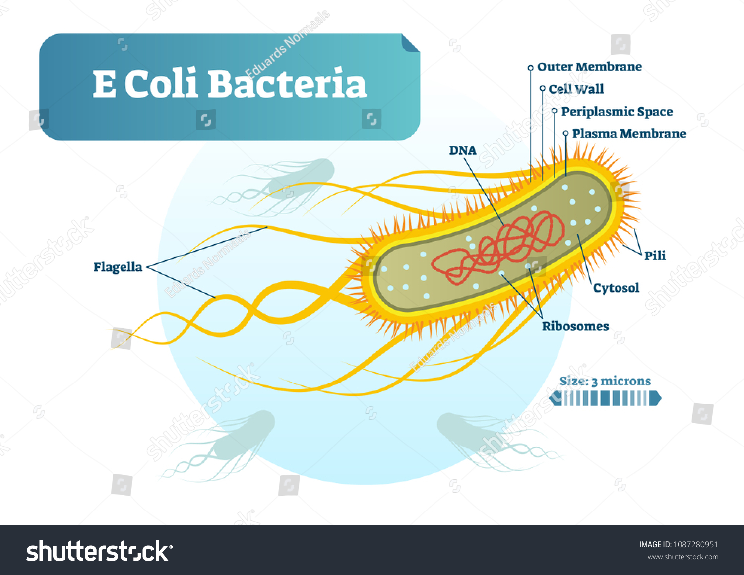


E Coli Bacteria Micro Biological Vector Stock Vector Royalty Free


Transmission Electron Microscopy Tem Micrographs Of E Coli K12 Nr50 Download Scientific Diagram
479 KB Escherichia coli flagella TEMpng 600 × 6;Escherichia coli Four different strains of Escherichia coli on Endo agar with biochemical slope Glucose fermentation with gas production, urea and H 2 S negative, lactose positive (with exception of strain D "late lactose fermenter";Escherichia coli bacteria on blood agar e coli under a microscope stock pictures, royaltyfree photos & images petri dishes with culture media for sarscov2 diagnostics, test coronavirus covid19, microbiological analysis e coli under a microscope stock pictures, royaltyfree photos & images
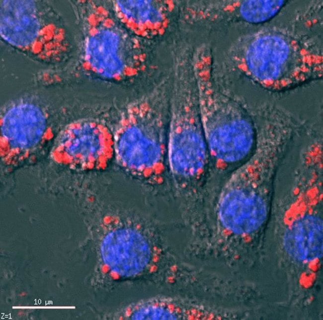


Phrodo Red I E Coli I Bioparticles Conjugate For Phagocytosis


Method For Labeling Transcripts In Individual Escherichia Coli Cells For Single Molecule Fluorescence In Situ Hybridization Experiments Protocol
Escherichia coli or Ecoli is a gramnegative species of bacillus shaped bacteria that can be easily observed under a microscope, even for those with the untrained eye This bacteria has a fast growth rate, doubling every minutes, making them a common choice for bacterial related research purposes• 1 Vial of E coli • 1 Microscope • 1 Vial of Immersion oil • 1 Vial of crystal violet dye • 1 Vial of Gram's Iodine • 1 Vial of Decolorizer • 1 Vial of Safranin dye • 13 Viewing slides • 1 Container of Lysol wipes 4 PreLab PreparationE coli as seen under an electron microscope What is microscope slide?


Bacterial Flagella Disrupt Host Cell Membranes And Interact With Cytoskeletal Components Biorxiv


Label Microscope Diagram Clipart Best
2 Observe the "e" under scanning, low and high power Draw or take a photomicrograph (depending on which version of the lab report you are completing) of what you see at 100xTM and 400xTM and label with the magnification power (Note Always label micrographs with the total magnifying power) 3Bacteria species E coli and S aureus under the microscope with different magnifications Bacteria are among the smallest, simplest and most ancient living• Label (Ex/Em) Fluorescein (~494/518 nm) • Particle E coli (K12 strain) • Opsonizing reagent available Using BioParticles Products BioParticles® conjugates are provided as lyophilized powders There are approximately 3 x 10⁸ E coli or S aureus particles per mg solid and approximately 2 x 10⁷ zymosan particles per mg solid BioParticles® conjugates can be reconstituted in the buffer of your choice for use in phagocytosis assays


What Does An E Coli Bacteria Look Like Under A Microscope Quora
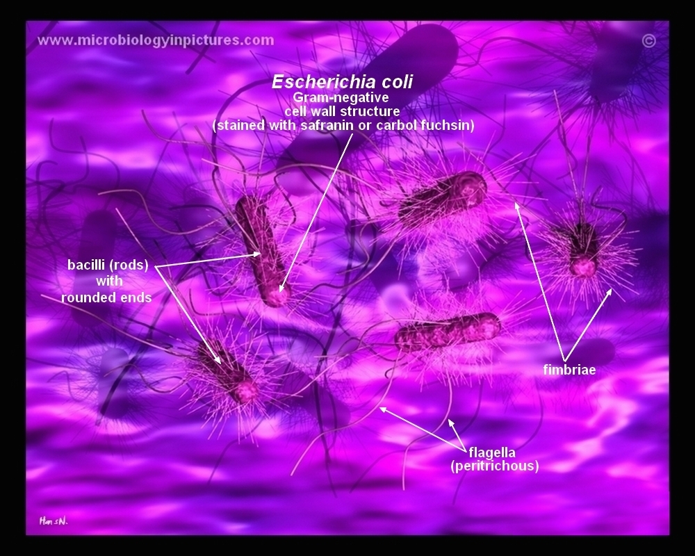


How E Coli Bacteria Look Like
Escherichia coli (/ ˌ ɛ ʃ ə ˈ r ɪ k i ə ˈ k oʊ l aɪ /), also known as E coli (/ ˌ iː ˈ k oʊ l aɪ /), is a Gramnegative, facultative anaerobic, rodshaped, coliform bacterium of the genus Escherichia that is commonly found in the lower intestine of warmblooded organisms (endotherms) Most E coli strains are harmless, but some serotypes (EPEC, ETEC etc) can cause serious food263 MB Escherichia coli electron microscopyjpg 800 × 640;E coli bacteria looks like small rods under compound microscope at 1000 magnification power It is gram negative bacterium which appears pink when stained by a staining technique called as Gram staining which is a differential staining technique 1K views


Human Skin Under Microscope Labeled Confocal Mosaicing Microscopy Of Human Skin Ex Vivo Spectral Photo Human Skin Human Anatomy Chart Microscopic Photography


Electron Microscopy Images As Well As A Sketch Of The E Coli And The Download Scientific Diagram
E Coli Under The Microscope Types Techniques Gram Stain Rapid Discrimination Of Gram Positive And Gram Negative Bacteria E Coli Under Microscope 400x Gram Stain For Films Sigma Aldrich Pin On Pursuit Diagram Compound Light Microscope Parts Labeled Microscope Stages Of Mitosis PicturesWith emission maxima at 509 and 5 nm respectively, EGFP and DsRed are suited for almost crossover free dual color labeling upon simultaneous excitation We imaged mixed populations of Escherichia coli expressing either EGFP or DsRed by onephoton confocal and by twophoton microscopy Both excitation modes proved to be suitable for imaging cells expressing either of the fluorescent proteinsA microscope slide is a very thin piece of glass used to hold the object that is to be viewed They usually have another smaller, very thin piece of glass called a coverslip which fits on top of the sample Labelled Diagram of a Microscope Remember Take care not to let
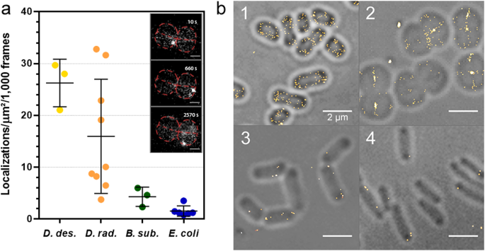


Bacterial Cell Wall Nanoimaging By Autoblinking Microscopy Scientific Reports


Single Molecule Imaging Of Electroporated Dye Labelled Chey In Live Escherichia Coli Philosophical Transactions Of The Royal Society B Biological Sciences
Yes, most of the bacteria range from 022 µm in diameter The length can range from 110 µm for filamentous or rodshaped bacteria The most wellknown bacteria E coli, their average size is ~15 µm in diameter and 26 µm in length As we talked above, you can see some bacteria in my cheek cellsEscherichia coli (E coli) bacteria normally live in the intestines of people and animals Most E coli are harmless and actually are an important part of a healthy human intestinal tract However, some E coli are pathogenic, meaning they can cause illness, either diarrhea or illness outside of the intestinal tract The types of E coli that can cause diarrhea can be transmitted throughEscherichia coli (/ ˌ ɛ ʃ ə ˈ r ɪ k i ə ˈ k oʊ l aɪ /), also known as E coli (/ ˌ iː ˈ k oʊ l aɪ /), is a Gramnegative, facultative anaerobic, rodshaped, coliform bacterium of the genus Escherichia that is commonly found in the lower intestine of warmblooded organisms (endotherms) Most E coli strains are harmless, but some serotypes (EPEC, ETEC etc) can cause serious food


Method For Labeling Transcripts In Individual Escherichia Coli Cells For Single Molecule Fluorescence In Situ Hybridization Experiments Protocol


Tata Complexes Exhibit A Marked Change In Organisation In Response To Expression Of The Tatbc Complex Biorxiv
E coli bacteria looks like small rods under compound microscope at 1000 magnification power It is gram negative bacterium which appears pink when stained by a staining technique called as Gram staining which is a differential staining technique 1K viewsAntigen 43 (Ag43) is a surfacedisplayed autotransporter protein of Escherichia coli By virtue of its selfassociation characteristics, this protein is able to mediate autoaggregation and flocculation of E coli cells in static cultures Additionally, surface display of Ag43 is associated with a distinct frizzy colony morphology in E coliSalmonella under the Microscope Salmonella is a genus of rodshaped bacteria found worldwide in both coldblooded and warmblooded animals as well as in the environment Strains of Salmonella cause illness such as typhoid fever, paratyphoid fever and food poisoning
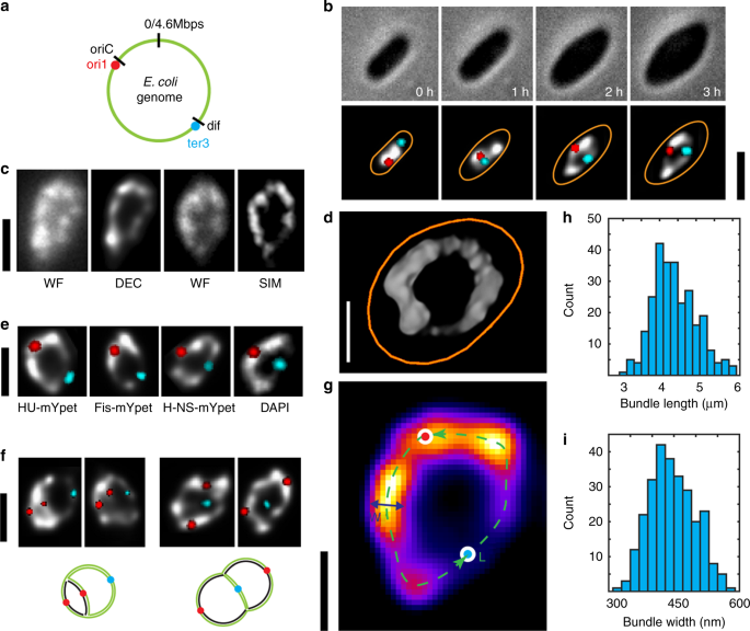


Direct Imaging Of The Circular Chromosome In A Live Bacterium Nature Communications


Staining Microscopic Specimens Microbiology
Luminiferous E coli B E cells, stained with the GFP‐labeled phage, were observed under an epifluorescence microscope (BX60, Olympus Co, Tokyo, Japan) equipped with an appropriate filter (U‐MWIBA/GFP, Olympus) Photographs were taken by cooled charge‐coupled digital camera (DP70, Olympus, Japan) under light and fluorescent fieldsE coli K12 cells could be detected under these crucial conditions, even in the absence of cultivation Download Download fullsize image;Escherichia coli and Staph aureus ()jpg 2,160 × 1,440;


Advantages Of Using Escherichia Coli As A Model Organism Andor Learning Centre Oxford Instruments


Ib Biology Notes 2 2 Prokaryotic Cells
E Coli E Coli under the microscope at 400x E Coli (Escherichia Coli) is a gramnegative, rodshaped bacterium Most E Coli strains are harmless, but some serotypes can cause food poisoning in their hosts The harmless strains are part of the normal flora of the gut Learn more about E Coli here Helicobacter PyloriThe nucleoid of living and OsO4 or glutaraldehydefixed cells of Escherichia coli strains was studied with a phasecontrast microscope, a confocal scanning light microscope, and an electron microscope The trustworthiness of the images obtained with the confocal scanning light microscope was investigated by comparison with phasecontrast micrographs and reconstructions based on seriallyNOTE If the chosen microscope does not have phase contrast or differential interference contrast capabilities, E coli outer membranes can be labeled with 2 μM lipophilic fluorescent marker (see table of materials), which should be added to the bacterial suspension min prior to fixation 17 Fixation and further treatment are accomplished


Frontiers A Fluorescence Based Proliferation Assay For The Identification Of Replicating Bacteria Within Host Cells Microbiology


E Coli Parts Page 1 Line 17qq Com
E coli are rodshaped bacterium that has an outer membrane consisting of lipopolysaccharides, inner cytoplasmic membrane, peptidoglycan layer, and an inner, cytoplasmic membrane Cell wall this structure is made of a thick layer of protein and sugar that prevents the cell from burstingE Coli Under The Microscope Types Techniques Gram Stain Ecoli Microscope 100x Oil Stock Photo Edit Now Individual Escherichia Coli Smear Prepared Glass Microscope Escherichia Coli Bacteria By Scanning Electron Microscopy E Coli Microscope Labelled Diagram GcseExamples of Bacteria Under the Microscope Escherichia coli Escherichia coli (Ecoli) is a common gramnegative bacterial species that is often one of the first ones to be observed by students Most strains of Ecoli are harmless to humans, but some are pathogens and are responsible for gastrointestinal infections They are a bacillus shaped bacteria that has a very fast growth (they can double every minutes), which is one of the main reasons they are used in research


Bacterial Flagella Disrupt Host Cell Membranes And Interact With Cytoskeletal Components Biorxiv
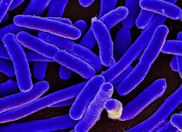


E Coli Under The Microscope Types Techniques Gram Stain Hanging Drop Method
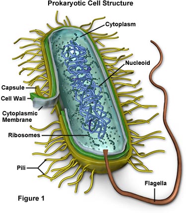


Molecular Expressions Cell Biology Bacteria Cell Structure


Structure And Function Of Bacterial Cells
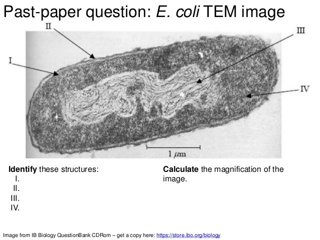


Ib Biology 1 2 Slides Ultrastructure Of Cells


The Interaction Of Escherichia Coli O157 H7 And Salmonella Typhimurium Flagella With Host Cell Membranes And Cytoskeletal Components Microbiology Society


Cardiolipin Microdomains Localize To Negatively Curved Regions Of Escherichia Coli Membranes Pnas


Imaging Host Pathogen Interactions Using Epithelial And Bacterial Cell Infection Models Journal Of Cell Science


Correlative Microscopy For Structural Microbiology Sciencedirect


Biol 230 Lab Manual Lab 1


Escherichia Coli Light Microscopy
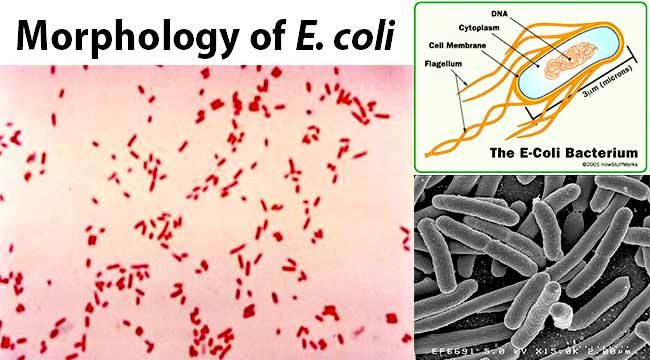


Escherichia Coli E Coli An Overview Microbe Notes


Iopscience Iop Org Article 10 10 1361 6463 f255 Pdf


Lab Manual Exercise 1


Light Microscope Images Of Filamentous E Coli Cells And Download Scientific Diagram


Bacteria Under The Microscope E Coli And S Aureus Youtube
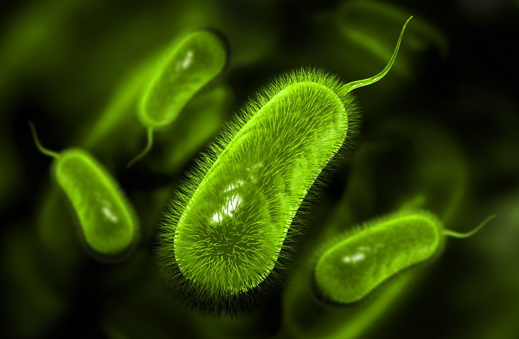


Bactag Gram Negative Bacterial Label The Native Antigen Company


The Five Major Groups Of Microbes


Optofluidic Label Free Sers Platform For Rapid Bacteria Detection In Serum Sciencedirect


Morphology Of E Coli Cells Under Microscope At 100 Magnification Download Scientific Diagram


O Tsutsugamushi Labelled With Peptidoglycan Reactive Probes A Download Scientific Diagram


Fluorescent Antibiotics New Research Tools To Fight Antibiotic Resistance Trends In Biotechnology


Iopscience Iop Org Article 10 10 1361 6463 f255 Pdf


Q Tbn And9gctffwwm42evo5c26svvkxccrastvfjru302ru 63nxjqythb6cl Usqp Cau


Live Cell Fluorescence Microscopy To Observe Essential Processes During Microbial Cell Growth Abstract Europe Pmc


Bactoview Live Fluorescent Bacterial Stains Biotium


Magnification Bioninja


Entamoeba Coli Wikipedia


Studying Age Associated Pathologies Through Temperature Controlled Microscopy Linkam Scientific


Structure And Function Of Bacterial Cells


Pdf Cell Segmentation By Multi Resolution Analysis And Maximum Likelihood Estimation Mamle


3


Vector Clipart E Coli Bacteria Micro Biological Vector Illustration Cross Section Labeled Diagram Medical Research Information Poster Vector Illustration Gg Gograph


A Microscopy Images Depicting Phagocytosis Of Labelled E Coli Download Scientific Diagram
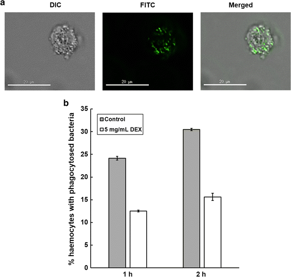


Effective Immunosuppression With Dexamethasone Phosphate In The Galleria Mellonella Larva Infection Model Resulting In Enhanced Virulence Of Escherichia Coli And Klebsiella Pneumoniae Springerlink


Confocal Fluorescent Microscopy Of Live E Coli Ag102 Labelled With Download Scientific Diagram
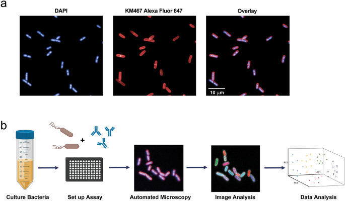


A Novel Therapeutic Antibody Screening Method Using Bacterial High Content Imaging Reveals Functional Antibody Binding Phenotypes Of Escherichia Coli St131 Scientific Reports


Unique Characteristics Of Prokaryotic Cells Microbiology


Phagocytosis Assay Kit Green E Coli Ab Abcam


2 2 Prokaryotic Cells Bioninja


2 2 3 Identify Structures In Electron Micrographs Of Ecoli Youtube
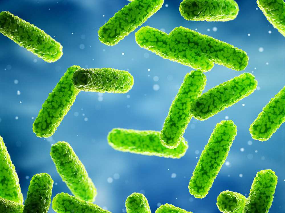


Legiotag Legionella Pneumophila Specific The Native Antigen Company


Staining Microscopic Specimens Microbiology


Q Tbn And9gcrxsv6valktvydnaiqm6 Yoim4dbn 9td0atqk2azvfczn73wcq Usqp Cau


Unique Characteristics Of Prokaryotic Cells Microbiology
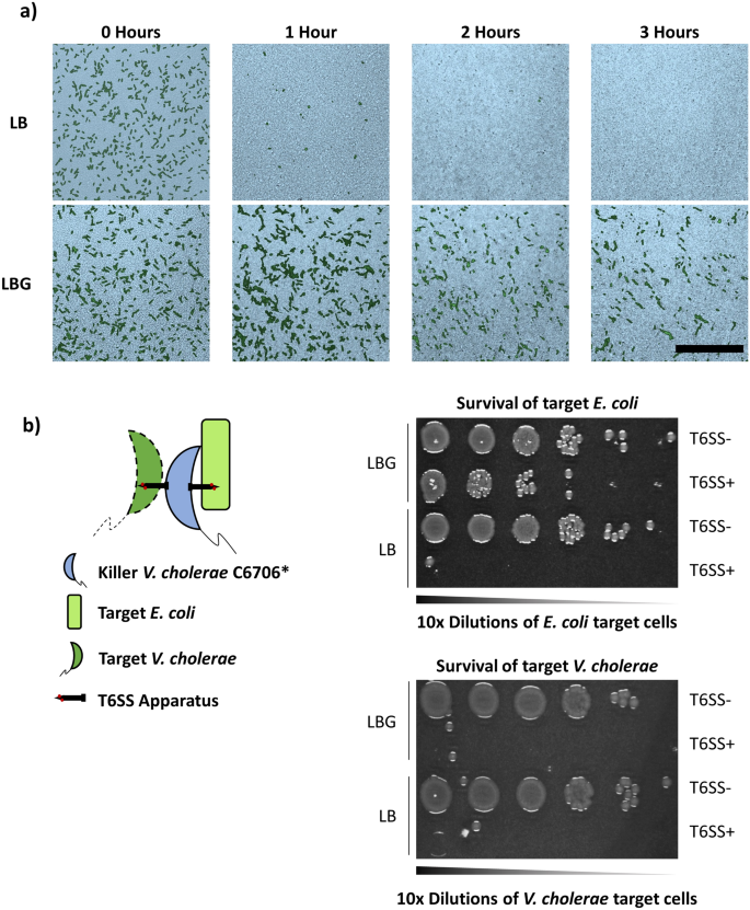


Glucose Confers Protection To Escherichia Coli Against Contact Killing By Vibrio Cholerae Scientific Reports
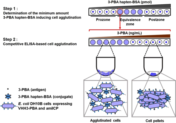


A Label Free Optical Whole Cell Escherichia Coli Biosensor For The Detection Of Pyrethroid Insecticide Exposure Scientific Reports
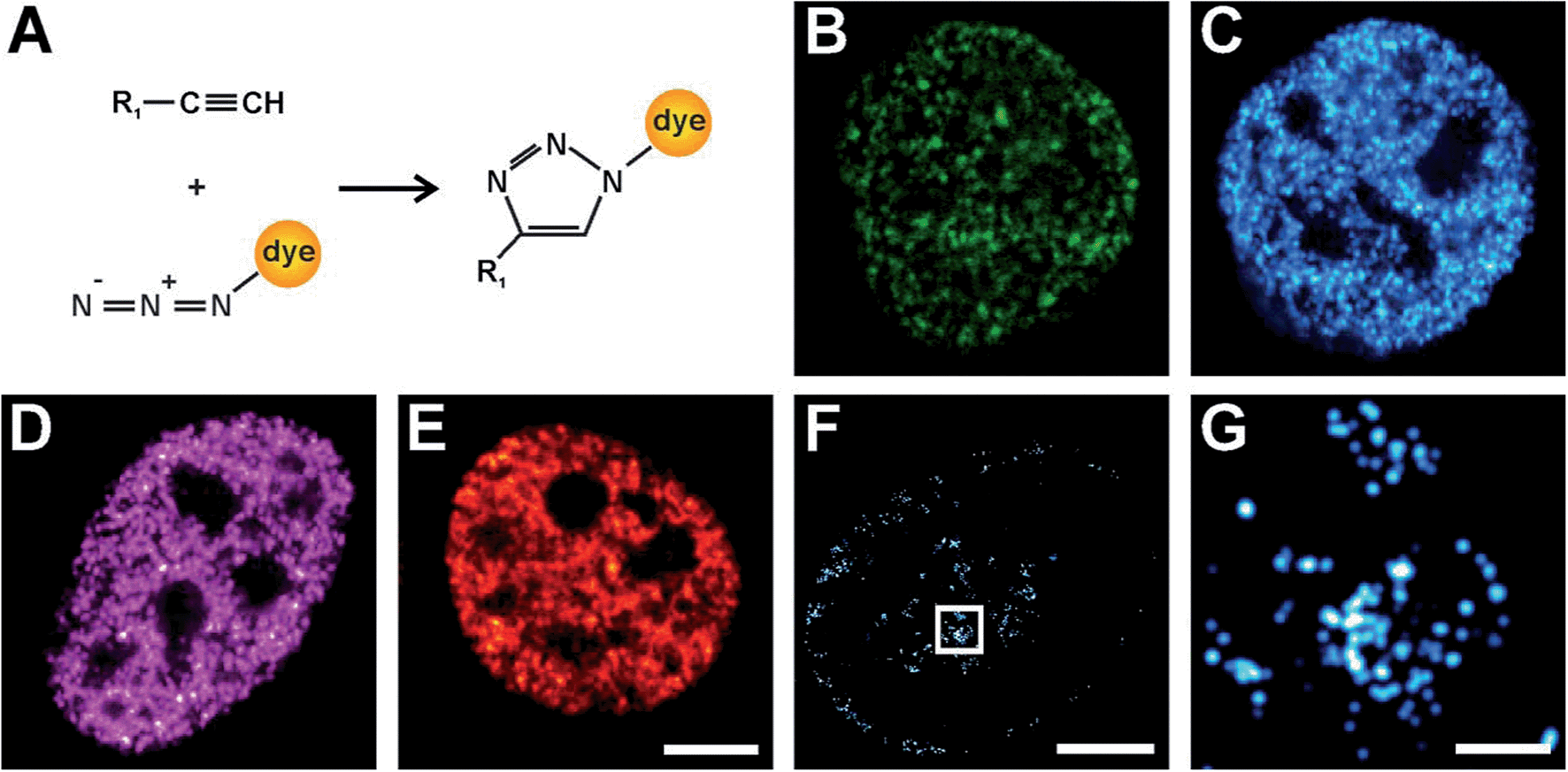


Click Chemistry Facilitates Direct Labelling And Super Resolution Imaging Of Nucleic Acids And Proteins Rsc Advances Rsc Publishing Doi 10 1039 C4rab
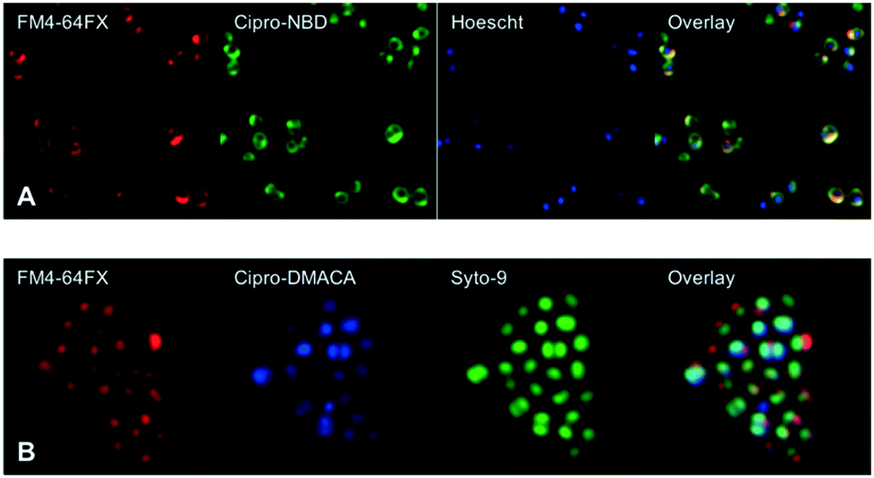


Fluoroquinolone Derived Fluorescent Probes For Studies Of Bacterial Penetration And Efflux Medchemcomm Rsc Publishing Doi 10 1039 C9mdg


Phrodo Red I E Coli I Bioparticles Conjugate For Phagocytosis
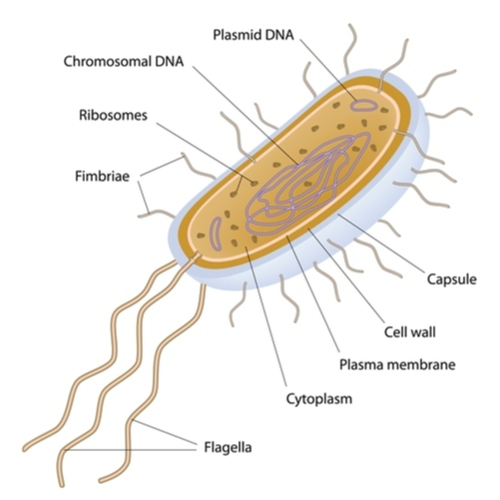


The Five Major Groups Of Microbes
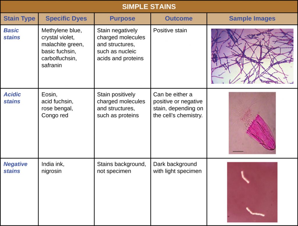


2 4 Staining Microscopic Specimens Microbiology Canadian Edition


Tel Archives Ouvertes Fr Tel Document
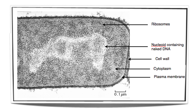


Ib Biology Notes 2 2 Prokaryotic Cells


Bacterial Characteristics Gram Staining Video Khan Academy


What Does An E Coli Bacteria Look Like Under A Microscope Quora
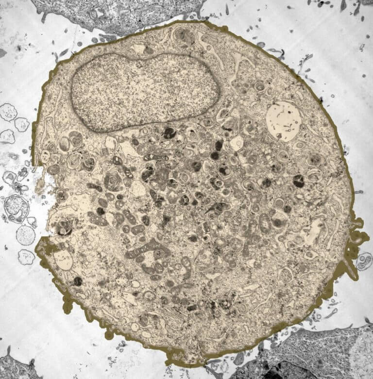


How These 26 Things Look Like Under The Microscope With Diagrams


Metabolic Labeling Of Escherichia Coli Genomic Dna With Erythrosine 11 Dutp For Functional Imaging Via Correlative Microscopy Loukanov Microscopy Research And Technique Wiley Online Library


Figure 1 From Supplementary Figure 1 Electron Microscopy And Diffraction Data Of Ompg Electron Microscopy Of Different Isotopically Labelled Ompg Preparations After Reconstitution Into E Coli Lipids And Growth Of 2d Crystals


Bacteria Cell Under Microscope Labeled


Representative Brightfield And Fluorescence Microscopy Images Of E Download Scientific Diagram


Fluorescent Antibody Techniques Microbiology


Segmentation Comparison Result Fluorescent Protein Lab Open I
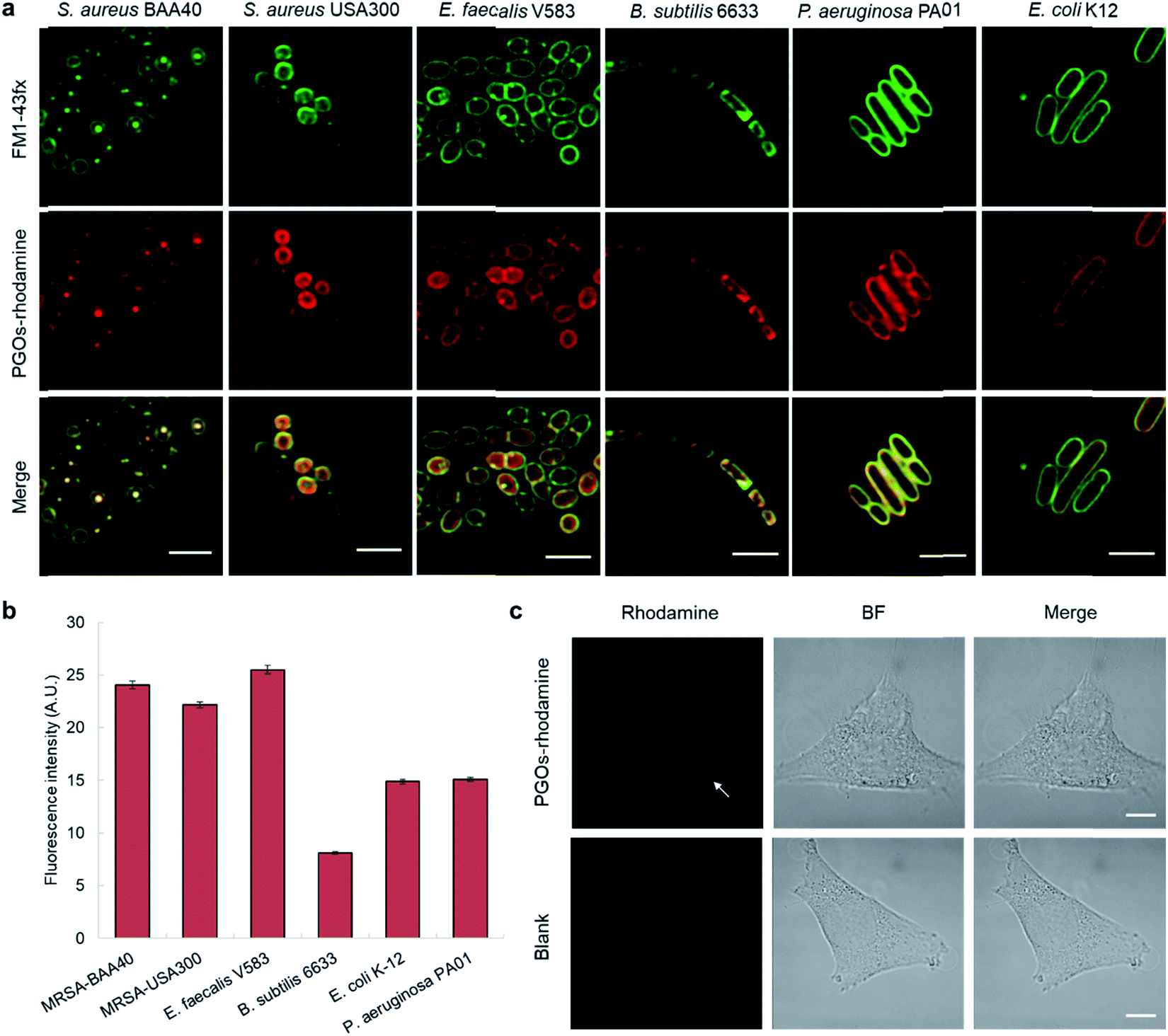


Synthetic Biohybrid Peptidoglycan Oligomers Enable Pan Bacteria Specific Labeling And Imaging In Vitro And In Vivo Chemical Science Rsc Publishing Doi 10 1039 C9sce
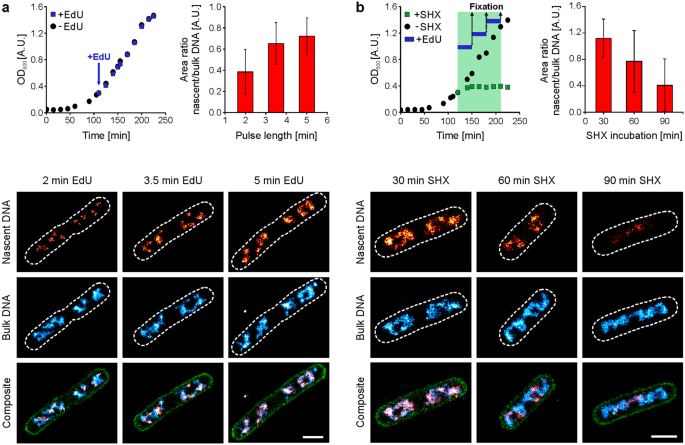


A Toolbox For Multiplexed Super Resolution Imaging Of The E Coli Nucleoid And Membrane Using Novel Paint Labels Scientific Reports


Tata Complexes Exhibit A Marked Change In Organisation In Response To Expression Of The Tatbc Complex Biorxiv


2 2 Prokaryotic Cells Bioninja


Confocal Microscopy Imaging Of Cells Expressing Different Markers And Download Scientific Diagram


Microbiology Stains Kits Dyes Bacteria Yeast Viability Pcr Biotium


Mechanisms Underlying Predatory Bacteria Revealed By Researchers Microbiology


What Does An E Coli Bacteria Look Like Under A Microscope Quora


What Does An E Coli Bacteria Look Like Under A Microscope Quora
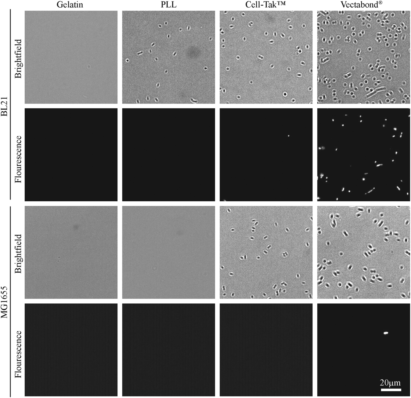


Imaging Live Bacteria At The Nanoscale Comparison Of Immobilisation Strategies Analyst Rsc Publishing Doi 10 1039 C9and
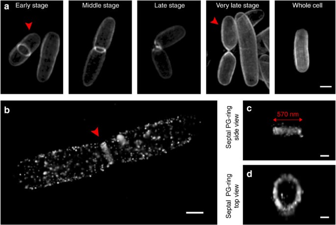


Metabolic Labelling Of The Carbohydrate Core In Bacterial Peptidoglycan And Its Applications Nature Communications


コメント
コメントを投稿